
What to Know About the Cardiac Catheterization Procedure
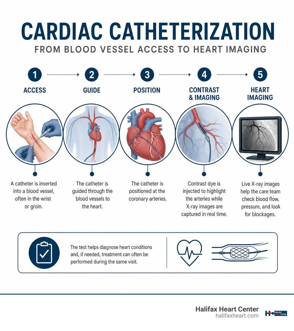
A cardiac catheterization procedure is a minimally invasive test that lets a heart specialist check blood flow, pressure, and possible blockages in and around the heart. A thin, flexible tube called a catheter is guided through a blood vessel, often in the wrist or groin, to the heart while the care team uses live X-ray imaging.
Most people are awake with local numbing medicine and light sedation. The test can help explain chest pain, shortness of breath, fatigue, or an irregular heartbeat – and, in some cases, a blockage can be treated during the same visit with angioplasty or a stent.
Cardiac catheterization is one of the most common heart procedures in the U.S. A diagnostic test often takes about 30 to 45 minutes, although preparation, recovery, and any treatment can make the visit longer.
At Halifax Heart Center, we provide patient-centered cardiovascular care for people across South Boston, Halifax County, and surrounding Southside Virginia communities. Our team helps patients understand each step of a cardiac catheterization procedure, from accurate diagnosis through personalized next-step care.
Cardiac catheterization procedure terms explained:
What Is a Cardiac Catheterization Procedure and How Does It Work?
A cardiac catheterization procedure, often referred to simply as a “heart cath,” is a specialized medical evaluation performed inside a dedicated cardiac catheterization laboratory (cath lab). During the test, an interventional cardiologist inserts a hollow, flexible plastic tube (catheter) into a blood vessel in your arm, wrist, or groin. Using advanced fluoroscopy guidance (a continuous X-ray movie), the physician gently maneuvers the catheter through your vascular system until it reaches the cardiac chambers and coronary arteries.
Once the catheter is positioned, the medical team injects a small amount of iodinated contrast dye into the vessels. Because this dye shows up clearly on X-ray imaging, it creates high-definition roadmaps of your coronary arteries, revealing areas of plaque buildup, narrowing, or complete blockage.
Beyond taking pictures of blood vessels, a diagnostic cardiac catheterization provides real-time intracardiac pressure measurements and hemodynamic monitoring. It is the only direct method medical professionals have to measure exact pressures inside every heart chamber and assess how efficiently blood flows through your valves and into the lungs.
Left Heart vs. Right Heart Catheterization
Depending on your symptoms and the specific questions your cardiologist needs answered, catheterization can evaluate the left side of your heart, the right side, or both.
| Feature | Left Heart Catheterization | Right Heart Catheterization |
|---|---|---|
| Primary Vascular Access | Arterial (Radial artery in wrist or Femoral artery in groin) | Venous (Femoral vein, internal jugular vein, or arm vein) |
| Target Anatomy | Aortic root, coronary arteries, left ventricle | Superior/inferior vena cava, right atrium, right ventricle, pulmonary arteries |
| Main Clinical Focus | Evaluating coronary artery blockages and left ventricular pumping strength | Measuring pulmonary pressures, cardiac output, and heart failure hemodynamics |
| Common Associated Tests | Selective coronary angiography, left ventriculography, FFR/iFR | Pulmonary capillary wedge pressure (PCWP), shunt runs, vasodilator challenges |
Following modern left heart catheterization protocols, arterial access allows doctors to engage the left main and right coronary arteries directly to check for flow-limiting obstructions. Right heart catheterization, by contrast, travels through systemic veins to calculate resting cardiac output (typically 4 to 8 L/min) and determine whether elevated pressures in the lungs (pulmonary hypertension) are contributing to symptoms like leg swelling and severe fatigue.
Advanced Diagnostic Tools Used During the Procedure
Modern cath labs utilize several cutting-edge diagnostic adjuncts that go beyond standard 2D X-ray images. When visual imaging shows a borderline narrowing (for example, a 50% to 70% blockage), your cardiologist can employ functional tools to decide whether the vessel truly needs a stent:
- Fractional Flow Reserve (FFR) & Non-Hyperemic Pressure Ratios (NHPR/iFR): A hair-thin sensor wire is placed across the narrowed artery to measure pressure drops. An FFR reading below 0.75 to 0.80 or an iFR of 0.89 or lower confirms that the blockage restricts blood flow enough to benefit from treatment.
- Intravascular Ultrasound (IVUS): A tiny ultrasound probe inside the catheter produces 360-degree cross-sectional images of the arterial wall, showing the composition of the plaque (soft cholesterol vs. hard calcium) and helping size stents precisely.
- Optical Coherence Tomography (OCT): Using light waves instead of sound waves, OCT provides microscopic imaging resolution of the vessel lining.
- Endomyocardial Biopsy: A specialized catheter with tiny, jaw-like forceps can harvest minute heart tissue samples to check for inflammatory cardiomyopathy or evaluate heart transplant health (cardiac perforation is an extremely rare risk, occurring in only 0.2% to 0.7% of biopsy cases).
These advanced techniques seamlessly complement noninvasive diagnostic heart testing such as echocardiograms and nuclear stress tests to give our clinical team an exact physiological picture.
Why Doctors Recommend Cardiac Catheterization
Doctors recommend a cardiac catheterization procedure when noninvasive tests show suspicious findings or when a patient experiences cardiovascular symptoms that require definitive answers. More than one million catheterizations are performed annually in the United States, making it a cornerstone of modern cardiac evaluation.
Common clinical reasons for catheterization include:
- Unexplained, persistent chest pain (angina) or chest tightness
- Shortness of breath during basic daily activities
- Inconclusive, abnormal, or high-risk findings on an exercise treadmill or nuclear stress test
- Unexplained heart failure or newly discovered reduced ejection fraction
- Evaluation of heart valve stenosis or regurgitation before surgical or transcatheter repair
- Monitoring for transplant organ rejection or ruling out complex congenital anomalies
Evaluating Coronary Artery Disease and Heart Symptoms
When plaque accumulates along the walls of the arteries that nourish the heart muscle (atherosclerosis), it reduces oxygen delivery, causing myocardial ischemia. A detailed cardiac catheterization overview from leading clinical registries highlights that angiography remains the undisputed gold standard for mapping this disease.
It helps identify single-vessel, multi-vessel, or critical left main disease, as well as microvascular angina (spasms or resistance in microscopic heart vessels that do not show up on basic scans). For patients across Southside Virginia experiencing progressive cardiovascular symptoms, our comprehensive heart services ensure that noninvasive evaluations and invasive diagnostic decisions are aligned with guideline-directed medical standards.
Interventions Performed During a Cardiac Catheterization Procedure
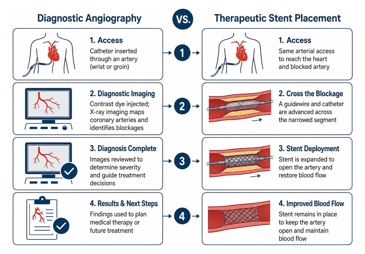
One of the greatest benefits of cardiac catheterization is its ability to transition immediately from a diagnostic evaluation into a therapeutic treatment during the very same session. If your interventional cardiologist finds a significant blockage, several minimally invasive options are available:
- Balloon Angioplasty: A catheter with an uninflated balloon at its tip is threaded across the blockage and gently inflated to compress plaque against the artery wall.
- Drug-Eluting Stent (DES) Placement: A small expandable metal mesh tube coated with medication is expanded at the site of the blockage. The medication slowly elutes into the artery wall to prevent scar tissue formation and re-narrowing (restenosis).
- Intravascular Lithotripsy (IVL) & Rotational Atherectomy: For heavily calcified, rigid plaque that cannot easily be dilated, sonic pressure waves (lithotripsy) or a microscopic diamond-tipped drill (atherectomy) can break up hard calcium deposits before stent deployment.
- Balloon Valvuloplasty & Structural Repairs: Catheters can widen stiff, narrowed heart valves or close structural holes like patent foramen ovale (PFO) and atrial septal defects (ASD) without requiring open-chest surgery.
Preparing for Your Catheterization: What to Expect Before and During
Preparing properly for a cardiac catheterization ensures your procedure goes smoothly and minimizes the risk of complications. Because mild conscious sedation is used to keep you relaxed and comfortable while awake, preparation starts well before you arrive at the facility.
Pre-Procedure Fasting and Medication Adjustments
Your care team will provide tailored instructions regarding which medications to continue or hold. Common pre-procedure protocols include:
- Fasting Instructions: Do not eat or drink anything (including water) for 6 to 8 hours prior to your scheduled procedure time.
- Blood Thinners (Anticoagulants): Medications such as warfarin, apixaban (Eliquis), rivaroxaban (Xarelto), or dabigatran (Pradaxa) are typically held 24 to 48 hours prior to elective catheterization depending on your kidney function and planned access site.
- Diabetes Medications: Oral diabetes medications are usually held on the morning of the procedure. If you take metformin, it must be held on the day of the procedure and for 48 hours afterward to prevent rare metabolic interactions with contrast dye.
- Contrast Dye Allergies: If you have had prior allergic reactions to radiographic contrast, shellfish, or iodine, inform your team in advance; a simple premedication regimen of oral prednisone and diphenhydramine (Benadryl) can prevent hypersensitivity reactions (severe reactions occur in only 0.02% to 0.04% of patients).
- Kidney Protection: Contrast dye is filtered by the kidneys. If you have underlying renal dysfunction, your team may administer intravenous normal saline hydration before and after the procedure to protect kidney health.
- Transportation: Because you will receive mild sedatives, you must arrange for a responsible adult friend or family member to drive you home after discharge.
Step-by-Step Breakdown of the Cardiac Catheterization Procedure
Knowing what happens during each phase of the test helps ease any natural apprehension:
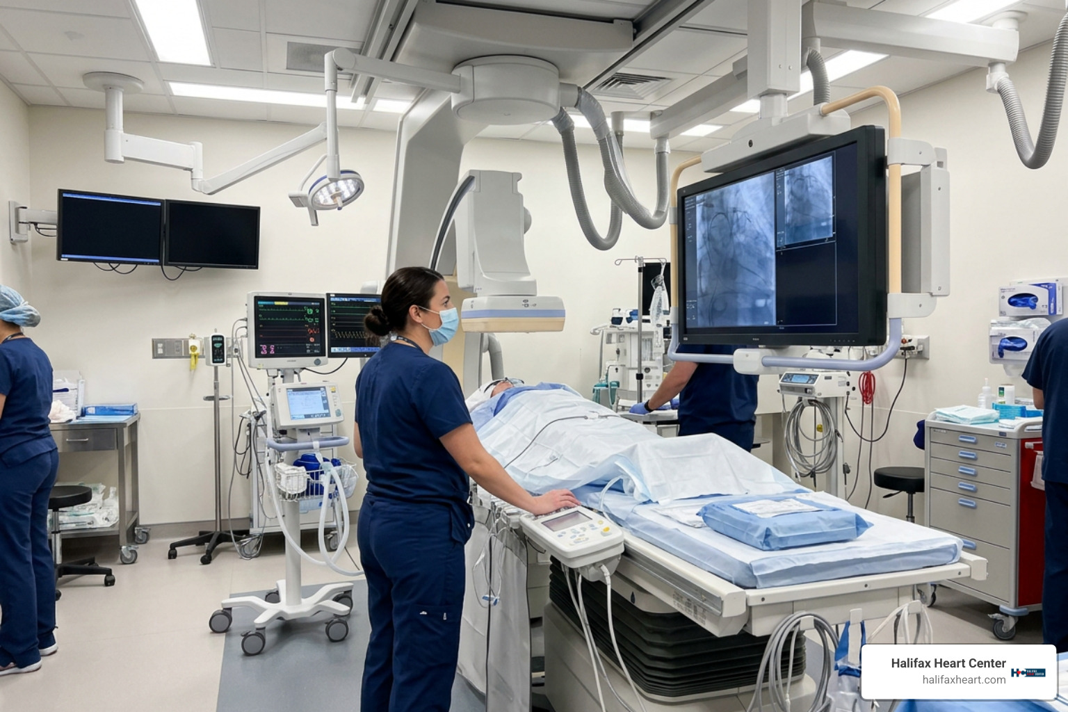
- Preparation and Monitoring: You will lie comfortably on a cushioned X-ray table. Cath lab nurses will place ECG sticky patches on your chest to continuously monitor your heart rate and rhythm, along with a blood pressure cuff and pulse oximeter.
- Sedation and Local Anesthesia: An IV line delivers mild relaxing medication. The medical team cleans your wrist (radial approach) or groin (femoral approach) with an antiseptic solution and injects a local numbing medication (lidocaine). You will feel a quick pinch and burn, followed by complete numbness in that spot.
- Sheath and Catheter Placement: The cardiologist inserts a small plastic tube called an introducer sheath into the artery using the Seldinger technique. The flexible diagnostic catheter is inserted through this sheath and navigated up to the heart. You will not feel the catheter moving through your bloodstream because blood vessel walls have no internal pain receptors.
- Contrast Injection and Angiography: As the catheter engages the coronary arteries, contrast dye is injected while X-ray cameras take pictures from various angles. During contrast injection, according to detailed coronary angiography details, you may experience a brief, warm flush or a mild metallic taste, which lasts only 10 to 15 seconds. If transient contrast-induced bradycardia occurs, the doctor may ask you to give a hearty cough, which clears the dye and normalizes your heart rate.
- Catheter Removal and Hemostasis: Once the diagnostic imaging and any necessary interventions are completed, the catheter and sheath are removed, and the insertion site is sealed to stop bleeding.
Recovery, Risks, and Understanding Your Results
After your procedure, you will be moved to the post-anesthesia recovery area for monitoring while your sedation wears off and the access puncture site stabilizes.
Radial vs. Femoral Access Recovery Guidelines
Recovery protocols depend heavily on whether your doctor used the radial artery (wrist) or femoral artery (groin). Current cardiovascular guidelines strongly favor radial-first access whenever anatomically feasible because it dramatically reduces bleeding complications and offers superior patient comfort.
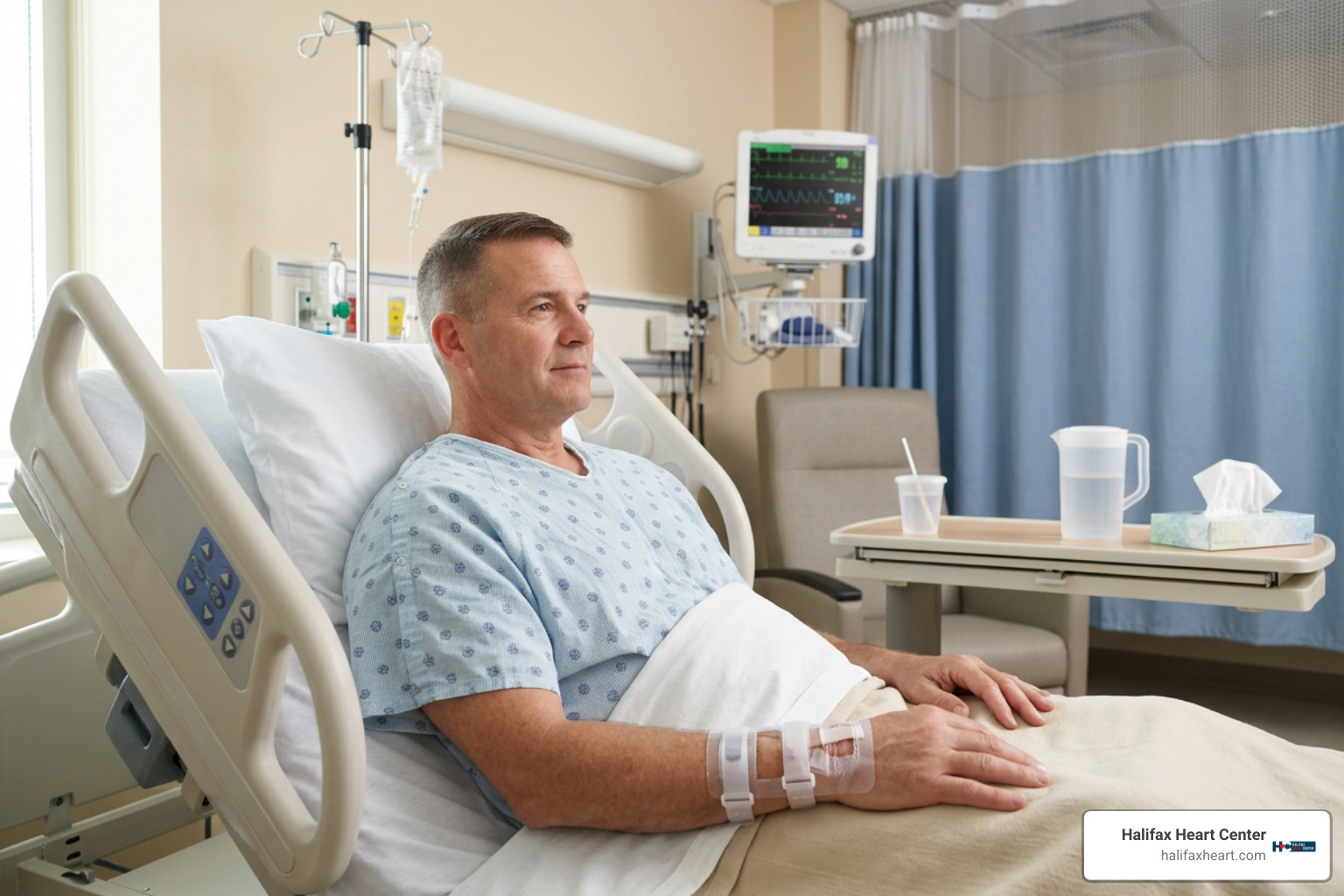
- Radial Access Recovery (Wrist): A clear pressure band (such as a TR Band) is placed over your wrist to compress the artery. You can generally sit up in bed immediately, enjoy a light meal, and walk within an hour. The wrist band is gradually deflated over 1 to 3 hours before discharge.
- Femoral Access Recovery (Groin): If the groin artery is used, hemostasis is achieved either by direct manual pressure for 15 to 20 minutes or by deploying a specialized vascular closure device (such as a collagen plug, clip, or suture). You must lie flat on your back with your leg kept completely straight for 2 to 6 hours to allow the artery to seal securely.
General At-Home Recovery Guidelines:
- Hydration: Drink several glasses of water over the next 24 hours to help your kidneys quickly flush out the contrast dye.
- Activity Resumption: Avoid strenuous physical exercise, running, and heavy lifting (nothing over 10 pounds) for 2 to 5 days.
- Incision Hygiene: You may shower 24 hours after your procedure. However, avoid submerging the puncture site in water (no bathtubs, hot tubs, or swimming pools) for 7 days until the skin has completely healed.
- Driving: Do not drive for at least 24 hours post-procedure to ensure all sedative medications have fully cleared your system.
Potential Risks and Complications
Diagnostic cardiac catheterization is a very safe procedure performed routinely across the country. The overall rate of major complications during a diagnostic catheterization is approximately 1%, with an associated mortality rate between 0.01% and 0.7%. In cases where percutaneous coronary interventions (angioplasty and stenting) are performed, the major complication rate is approximately 4.5%.
Minor side effects include small bruising, localized tenderness, or a small, firm pea-sized knot under the skin, which is normal and typically resolves within a few weeks.
Rare complications (<1%) include severe bleeding, arterial wall tears (dissection), access site pseudoaneurysm, blood clots, stroke (<0.1% in diagnostic cases), heart attack, or contrast-induced acute kidney injury.
Call our clinic or seek emergency medical care if you experience any of the following warning signs:
- Active, continuous bleeding or rapid, visible swelling at the catheter insertion site
- Severe, worsening pain, extreme redness, warmth, or drainage at the puncture site
- Coldness, pale color, numbness, or tingling in the hand, arm, leg, or foot below the puncture site
- Chest pain, tightness, or shortness of breath
- Dizziness, fainting, or sudden irregular heartbeats
- A fever over 101°F
(Emergency tip: If active bleeding occurs at home, lie flat immediately, apply firm, continuous pressure directly over the puncture site with clean gauze, and call 911 if bleeding does not stop after 10 minutes).
Interpreting Results and Next Steps in Treatment
Before you leave the recovery unit, your cardiologist will discuss your preliminary findings. These results provide an objective assessment of your cardiovascular system:
- Normal Findings: The coronary arteries are open with smooth walls, normal blood flow, and normal intracardiac pressures and ejection fraction.
- Non-Obstructive Plaque: Mild plaque buildup is visible (less than 50% luminal narrowing) that does not restrict blood flow. This finding emphasizes the importance of aggressive lifestyle management, lipid-lowering medications, and blood pressure control to prevent disease progression.
- Significant Stenosis (Blockage): Blockages narrowing the artery lumen by 70% or more (or 50% in the left main coronary artery) are clinically significant.
Depending on the number, location, and complexity of blockages, your next steps may include:
- Optimal Medical Therapy: Managing symptoms through antiplatelet medications, beta-blockers, statins, and ACE inhibitors.
- Completed Stenting: If a stent was placed during your cath, you will take dual antiplatelet therapy (such as aspirin combined with clopidogrel, ticagrelor, or prasugrel) to keep the stent open.
- Surgical Consultation (CABG): For severe multi-vessel blockages, left main disease, or complex anatomy in diabetic patients, coronary artery bypass graft (CABG) surgery may provide the safest long-term outcome.
- Cardiac Rehabilitation: Structured, medically supervised exercise and nutrition programs designed to strengthen your heart muscle.
Our team of experienced providers at Halifax Heart Center reviews these diagnostic metrics in detail with each patient, ensuring that treatment recommendations are clearly understood and tailored to your everyday lifestyle.
Frequently Asked Questions About Cardiac Catheterization
How long does a heart catheterization take?
A purely diagnostic cardiac catheterization procedure that evaluates coronary arteries and heart pressures typically takes about 30 to 45 minutes to complete. If an intervention like balloon angioplasty, atherectomy, or stent placement is performed, the procedure may take 1 to 2 hours. Including pre-procedure check-in, prep, and post-procedure recovery room observation, patients should expect their total hospital visit to span approximately 5 to 8 hours.
Is cardiac catheterization considered major surgery?
No, cardiac catheterization is not major surgery. It is a minimally invasive endovascular procedure performed through a pinhole puncture in a blood vessel without surgical incisions, general anesthesia, or breathing tubes. Most patients remain awake, relaxed under mild conscious sedation, and return home on the same day.
When can I resume normal activities and driving after the procedure?
You can typically resume light everyday walking and sedentary activities the day after your procedure. You should refrain from driving for at least 24 hours after receiving sedation. Heavy physical lifting (over 10 pounds), strenuous cardiovascular exercise, and vigorous sports should be avoided for 2 to 5 days to allow the puncture site to heal properly. Most patients with uncomplicated diagnostic catheterizations can return to desk work within 48 hours.
Conclusion
A cardiac catheterization procedure remains medicine’s most reliable and informative gold-standard diagnostic tool for evaluating coronary artery disease, heart valve function, and cardiac pressures. By understanding how the test works, what to expect during preparation, and how simple recovery can be—especially with modern wrist-access techniques—you can approach your appointment with peace of mind and complete confidence.
At Halifax Heart Center, we are dedicated to providing clear communication, compassionate diagnostic testing, and individualized cardiovascular care for our neighbors in South Boston, Halifax County, and across Southside Virginia. If you have been experiencing unexplained chest discomfort or shortness of breath, or if you need an expert cardiovascular evaluation, schedule your consultation for comprehensive heart disease diagnosis & treatment with our clinical team today.