
Cardiac Arrest Emergency Treatment Starts in the First Minutes
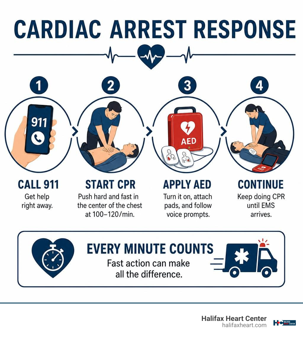
If someone suddenly collapses, is unresponsive, and is not breathing normally or is only gasping, call 911, start chest compressions, and use an AED as soon as one arrives. This is the most important cardiac arrest emergency treatment a bystander can provide while waiting for EMS.
- Make sure the area is safe.
- Call 911 on speakerphone, or point to someone and tell them to call and get an AED.
- Push hard and fast in the center of the chest at 100 to 120 compressions per minute.
- Turn on the AED, place the pads as shown, and follow its voice prompts.
- Keep doing CPR until the person starts moving or breathing normally, or emergency responders take over.
Cardiac arrest is an electrical failure that stops the heart from pumping blood. It is different from a heart attack, which is usually a blocked blood vessel. A heart attack can cause cardiac arrest, but a person having a heart attack may still be awake and breathing.
Time matters. Brain injury can begin within about five minutes without oxygen, and each minute without defibrillation can sharply lower the chance of survival. Gasping or snoring-like breaths are not normal breathing – treat them as a sign to begin CPR.
At Halifax Heart Center, we provide prevention, diagnostic testing, and personalized cardiovascular care for patients across South Boston, Halifax County, and Southside Virginia. While cardiac arrest emergency treatment begins with 911, CPR, and an AED, our team helps patients understand and manage the heart conditions that can raise cardiac risk.
Cardiac arrest emergency treatment terms at a glance:
- coronary artery disease treatment
- early signs of heart disease in females
- exercise prevents heart disease
Understanding Sudden Cardiac Arrest vs. Heart Attack
People often use the terms “cardiac arrest” and “heart attack” interchangeably, but they describe two distinct medical emergencies.
A heart attack (myocardial infarction) is primarily a plumbing problem. It occurs when a coronary artery becomes blocked, depriving a portion of the heart muscle of oxygen-rich blood. During a heart attack, the person is typically conscious, experiencing symptoms like chest pressure, shortness of breath, nausea, or cold sweats.
A sudden cardiac arrest is an electrical malfunction. The heart’s internal electrical signaling short-circuits, throwing the ventricles into a chaotic, uncoordinated flutter known as ventricular fibrillation (VF) or causing the heart’s mechanical pumping to stop altogether. Blood immediately ceases flowing to the brain, lungs, and vital organs. The individual collapses, loses consciousness within seconds, and will die within minutes without intervention.
While a heart attack can trigger cardiac arrest by destabilizing the heart’s electrical pathways, cardiac arrest can also result from structural heart conditions, severe electrolyte disturbances, inherited channelopathies, or physical trauma. It is also vital to understand how heart disease symptoms differ in women, as atypical warning signs can precede acute coronary events.
| Feature | Heart Attack (Myocardial Infarction) | Sudden Cardiac Arrest (SCA) |
|---|---|---|
| Primary Problem | Plumbing: Blood flow to the heart muscle is blocked | Electrical: The heart’s rhythm malfunctions abruptly |
| Consciousness | Usually awake, responsive, and speaking | Unresponsive and unconscious within seconds |
| Breathing | Breathing, though often labored or short of breath | Absent breathing or abnormal agonal gasping |
| Pulse | Present (may be rapid, slow, or irregular) | Absent |
| Primary Action | Call 911, keep calm, administer aspirin if advised | Call 911, start immediate CPR, use an AED |
Recognizing Immediate Signs and Warning Symptoms
Sudden cardiac arrest strikes without polite preamble. The most prominent signs include:
- Immediate collapse: The person falls limp without warning.
- Complete unresponsiveness: They do not react when you shout or tap their shoulders firmly.
- Absent or abnormal breathing: The person is either not breathing at all or demonstrating agonal respirations—irregular, labored snorts, moans, or gasps that occur when the respiratory center of the brain stem is starved of oxygen.
- Loss of pulse: Within seconds of arrest, systemic circulation plummets to zero.
When circulation stops, the cerebral hypoxia timeline begins:
- 0 to 10 seconds: The brain exhausts its residual oxygen reserves; loss of consciousness occurs.
- 5 minutes: Irreversible neuronal damage and brain injury can begin.
- 8 to 10 minutes: Survival rates drop precipitously without CPR.
Every single minute that passes without chest compressions and defibrillation reduces the chance of survival by 7% to 10%.
Step-by-Step Cardiac Arrest Emergency Treatment for Bystanders

When witnessing an adult collapse, bystanders must act swiftly. You do not need formal medical certification to save a life; you just need to take immediate, structured action.
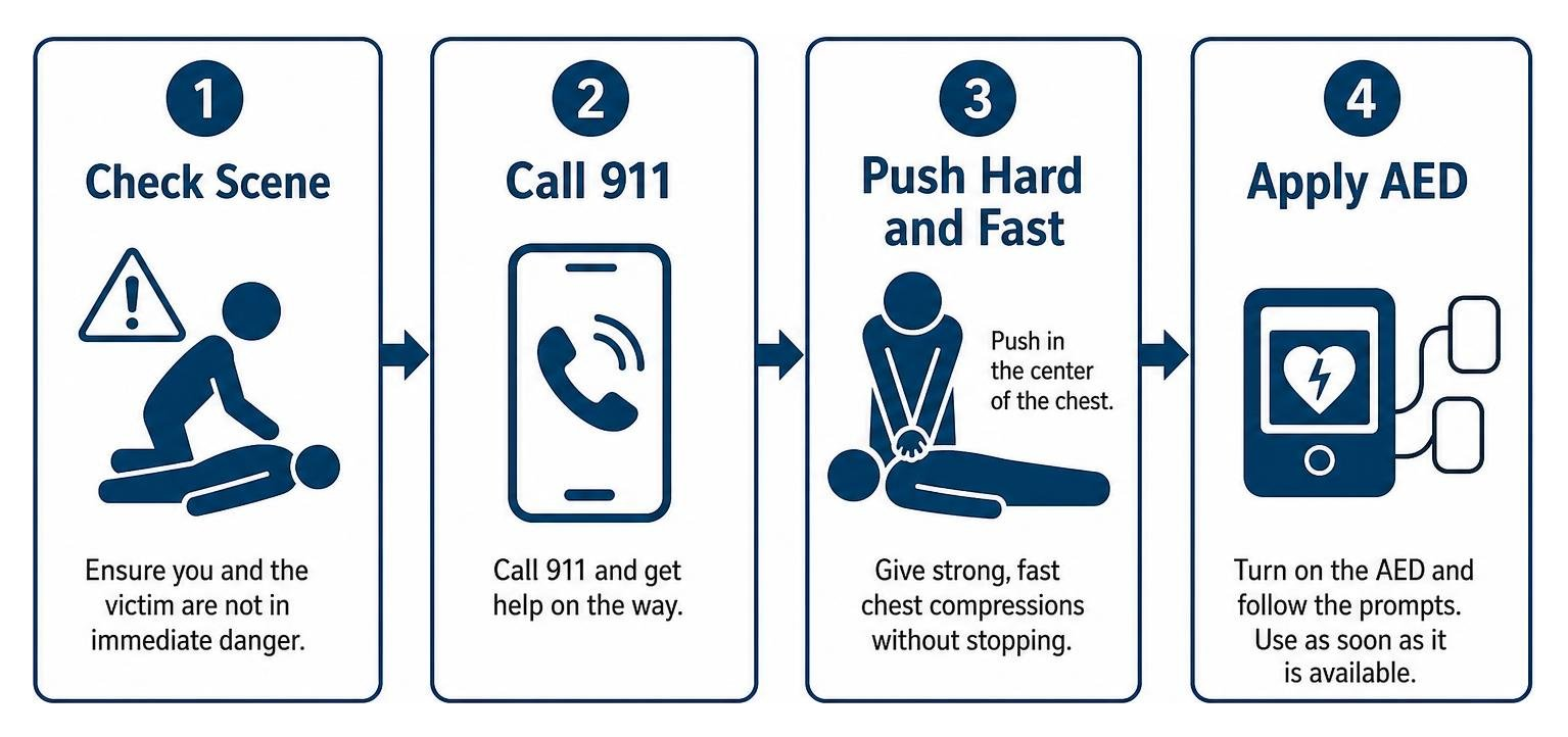
- Verify Scene Safety: Ensure you and the victim are not in immediate physical danger (such as active traffic, fire, or exposed electrical wires).
- Check for Responsiveness: Tap the victim’s shoulders firmly and shout, “Are you okay?” If there is no response, check their breathing. If they are not breathing or are only gasping, assume cardiac arrest.
- Activate Emergency Response (Call 911): If you are alone, call 911 on your mobile phone, activate the speakerphone, and place it next to you so you can talk to dispatch while assisting the person. If other people are present, point directly at one person and instruct: “You—call 911 and tell them someone collapsed, then find an AED!” When coordinating with Emergency Services | Halifax County, VA, clearly state your exact physical location, entrance, and landmark details.
- Begin Immediate Chest Compressions: Place the heel of one hand in the center of the chest and your other hand on top, interlacing your fingers. Push hard and fast.
- Deploy the AED: As soon as an AED arrives, open it, turn it on, and follow the voice and visual prompts without interrupting chest compressions any longer than necessary.
Immediate CPR Protocol in Cardiac Arrest Emergency Treatment
The foundation of resuscitation is high-quality cardiopulmonary resuscitation. By mechanically compressing the sternum against the spine, you artificially pressurize the cardiovascular system, forcing residual oxygenated blood through the coronary arteries to preserve the heart muscle and upward through the carotid arteries to sustain the brain.
To deliver high-quality CPR according to the Part 7: Adult Basic Life Support | American Heart Association CPR & First Aid guidelines:
- Rate: Push at a frequency of 100 to 120 compressions per minute (the tempo of the classic song “Stayin’ Alive” or “Baby Shark”).
- Depth: Compress the adult chest to a depth of at least 2 inches (5 to 6 cm), but no deeper than 2.4 inches.
- Full Recoil: Allow the chest to rise completely between compressions. Leaning on the chest prevents the heart chambers from refilling with blood, significantly degrading cardiac output.
- Hand Placement: Position the heel of your dominant hand on the lower half of the sternum (the center of the chest between the nipples), interlocking the fingers of your second hand to keep your fingertips lifted off the ribs.
- Minimize Interruptions: Keep hands-off time to fewer than 10 seconds. Every pause causes perfusion pressure in the brain and heart to crash back to baseline.
- Compression-to-Ventilation Ratio: If you are trained and willing to deliver rescue breaths, maintain a ratio of 30 compressions to 2 breaths. If you are untrained or prefer not to perform mouth-to-mouth, perform continuous Hands-Only CPR. Research shows that Hands-Only CPR is just as effective as traditional CPR with breaths during the first critical minutes of adult out-of-hospital cardiac arrest.
Deploying an Automated External Defibrillator (AED)
An Automated External Defibrillator (AED) is a sophisticated, computerized medical device designed to analyze the heart’s electrical rhythm and deliver a corrective electrical shock if a lethal rhythm is detected. Modern AEDs are engineered for absolute safety: they will never shock someone who does not need it.
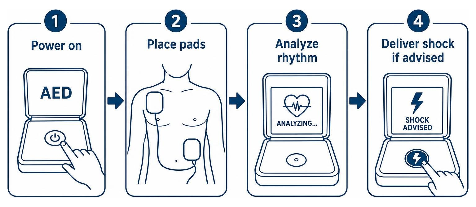
- Turn on the AED: Open the lid or press the power button. The unit will begin issuing spoken instructions.
- Expose and Prep the Chest: Bare the person’s chest. Wipe away excessive moisture or sweat with a cloth. If the individual has a medication patch on their chest, remove it with a gloved hand and wipe the skin clean.
- Apply the Adhesive Pads: Peel the backing off the pads and apply them firmly to the bare skin as illustrated on the packaging:
- Upper Right Pad: Placed directly below the right collarbone, to the right of the breastbone.
- Lower Left Pad: Placed on the lower left side of the ribs, a few inches below the armpit. (Note: If the person has a visible implanted pacemaker or defibrillator bump beneath the skin, position the pad at least 1 inch or 8 cm away from the device).
- Allow the AED to Analyze: Plug in the cable (if not pre-connected). When the device announces, “Analyzing heart rhythm, do not touch the patient,” shout “Clear!” and make sure no one is in contact with the individual.
- Deliver Shock if Advised: If the AED detects ventricular fibrillation or pulseless ventricular tachycardia, it will charge and instruct you to push the flashing shock button (fully automatic models will count down and deliver the shock automatically). Shout “Stand clear!” and confirm no one is touching the victim before the shock is administered.
- Resume CPR Immediately: Do not pause to check for a pulse or breathing after the shock. The AED will instruct you to resume chest compressions immediately for two minutes before it re-evaluates the heart rhythm.
Learning these steps builds lifesaving confidence. We encourage our community to explore opportunities like CPR for Healthcare Providers – EMS 100 at Southside Virginia … to practice hands-on resuscitation and AED deployment.
Clinical Resuscitation and Advanced Life Support Interventions
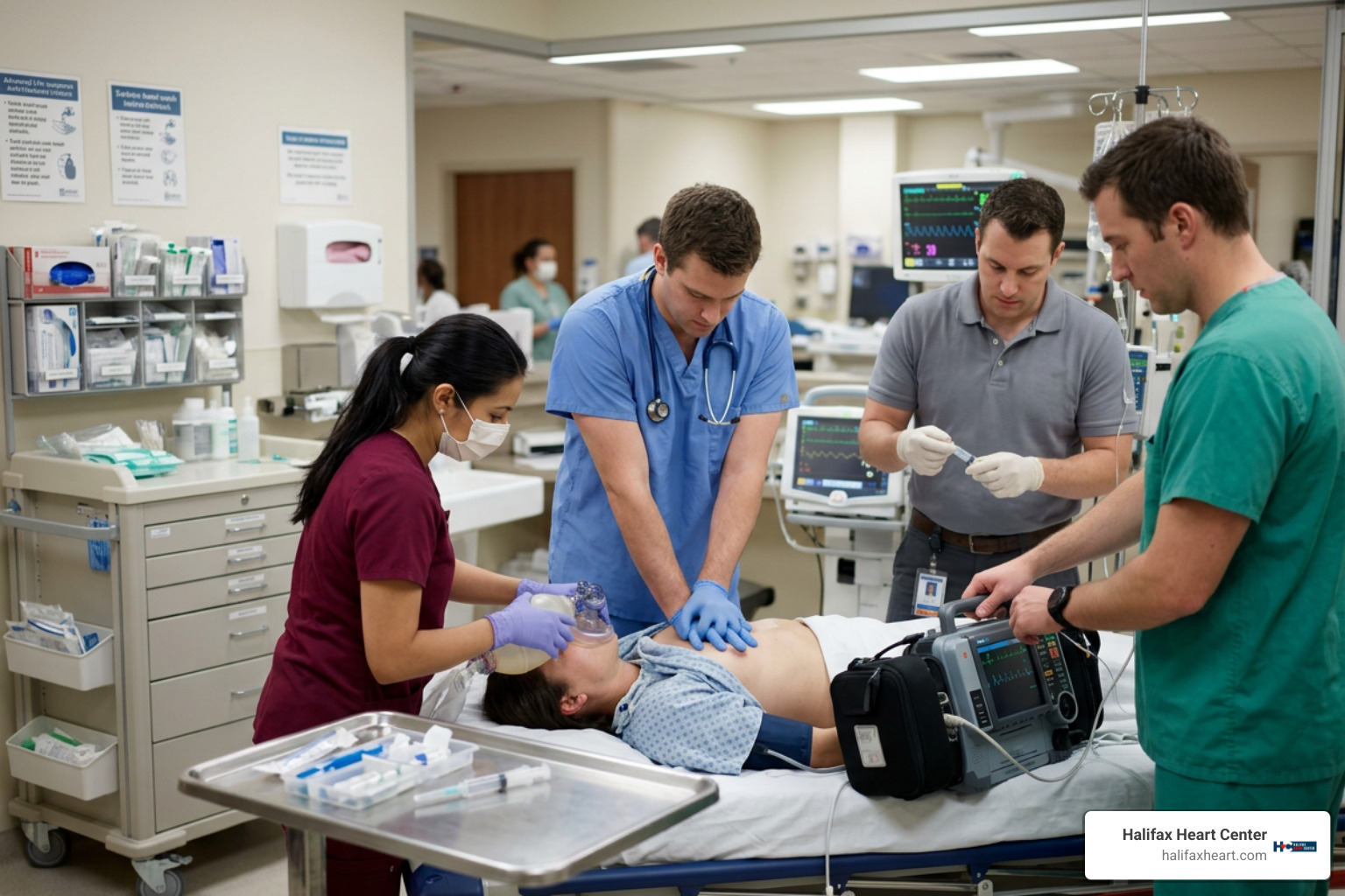
When emergency medical services (EMS) personnel and hospital emergency resuscitation teams arrive, basic life support transitions seamlessly into Advanced Cardiovascular Life Support (ACLS). Clinical responders utilize the Adult Cardiac Arrest Algorithm and standardized clinical protocols outlined in the Cardiopulmonary Resuscitation (CPR) in Adults – Critical Care Medicine – MSD Manual Professional Edition.
Resuscitation pathways diverge immediately based on whether the monitored cardiac rhythm is shockable or non-shockable:
- Shockable Rhythms (Ventricular Fibrillation & Pulseless Ventricular Tachycardia): Characterized by disorganized electrical firing. The immediate priority is early, unsynchronized biphasic defibrillation (120 to 200 Joules) combined with uninterrupted cycles of CPR and antiarrhythmic pharmacotherapy.
- Non-Shockable Rhythms (Asystole & Pulseless Electrical Activity – PEA): In asystole (“flatline”), there is a complete absence of electrical and mechanical activity. In PEA, organized electrical activity shows on the monitor, but the myocardium fails to contract effectively to generate a palpable pulse. Defibrillation does not work on these rhythms; the clinical focus shifts entirely to high-quality compressions, immediate vasopressor therapy, and diagnosing underlying reversible physiological causes.
Advanced Hospital and EMS Cardiac Arrest Emergency Treatment Protocols
Advanced life support protocols integrate continuous physiological monitoring, invasive airway management, and targeted pharmacotherapy:
- Vasopressor Therapy (Epinephrine): Standard ACLS dosing is 1 mg of Epinephrine (1:10,000 solution) administered IV/IO every 3 to 5 minutes. For non-shockable rhythms, guidelines mandate administering epinephrine as early as vascular access is established. For shockable rhythms, it is administered after initial defibrillation attempts have failed to terminate the arrhythmia.
- Antiarrhythmic Medications: For refractory ventricular fibrillation or pulseless VT persisting through initial shocks, clinicians administer Amiodarone (initial bolus of 300 mg IV/IO, followed by a second dose of 150 mg) or Lidocaine (1.0 to 1.5 mg/kg initial bolus).
- Airway & Waveform Capnography: Rescuers maintain oxygenation via bag-valve-mask or advance to supraglottic airway devices (such as the i-gel) or endotracheal intubation. Continuous quantitative end-tidal CO2 (ETCO2) waveform capnography is monitored. An ETCO2 value consistently below 10 mmHg indicates poor compression quality, while an abrupt, sustained spike in ETCO2 (typically >= 35–40 mmHg) is a primary indicator of the Return of Spontaneous Circulation (ROSC).
- Updated Guidelines Consensus: In alignment with the 2023 American Heart Association Focused Update on Adult Advanced Cardiovascular Life Support: An Update to the American Heart Association Guidelines for Cardiopulmonary Resuscitation and Emergency Cardiovascular Care and the European Resuscitation Council Guidelines 2025 Adult Advanced Life Support, the routine administration of calcium, magnesium, or sodium bicarbonate during undifferentiated cardiac arrest is no longer recommended unless specific clinical indications (such as hyperkalemia or calcium-channel blocker toxicity) are identified.
Identifying Reversible Causes: The H’s and T’s Framework
During active resuscitation, especially in non-shockable PEA and asystole arrests, the medical team systematically investigates and corrects the classic “H’s and T’s”:
- Hypovolemia: Severe blood or fluid loss; treated with rapid intravenous crystalloid fluids or blood product resuscitation.
- Hypoxia: Inadequate oxygen saturation; corrected with high-flow oxygenation and advanced airway placement.
- Hydrogen Ion (Acidosis): Severe metabolic acidosis; managed with effective ventilation and targeted buffer therapy.
- Hypo- / Hyperkalemia: Life-threatening potassium imbalances; hyperkalemia is stabilized with intravenous calcium gluconate/chloride, insulin with glucose, and sodium bicarbonate.
- Hypothermia: Core body temperature drops dangerously low; managed with active rewarming protocols.
- Tension Pneumothorax: Air trapped in the pleural cavity shifts mediastinal structures and blocks venous return; treated with immediate needle decompression or thoracostomy.
- Tamponade (Cardiac): Fluid accumulation in the pericardial sac constricts cardiac filling; relieved through emergency bedside pericardiocentesis.
- Toxins: Accidental or intentional overdoses (e.g., opioids, beta-blockers, tricyclic antidepressants); managed with specific reversal agents like naloxone and supportive antidotes.
- Thrombosis (Pulmonary Embolism): Massive blood clot occluding pulmonary vasculature; managed with emergency systemic thrombolytics or surgical embolectomy.
- Thrombosis (Coronary): Acute plaque rupture causing massive myocardial infarction; treated with immediate coronary reperfusion in the cardiac catheterization suite.
Post-Resuscitation Care and Long-Term Prevention
Achieving Return of Spontaneous Circulation (ROSC) is only the beginning of recovery. The post-cardiac arrest phase focuses on mitigating multi-organ injury and protecting the central nervous system from reperfusion injury.
- Targeted Temperature Management (TTM): For comatose survivors who do not follow verbal commands post-ROSC, clinicians implement targeted temperature control, continuously maintaining a stable core temperature between 32°C and 37.5°C for at least 24 to 72 hours. This suppresses harmful neuro-inflammatory cascades, prevents secondary cerebral edema, and avoids fever spikes.
- Hemodynamic and Respiratory Optimization: Blood pressure is tightly titrated using vasopressors to maintain a Mean Arterial Pressure (MAP) of at least 65 to 70 mmHg, avoiding hypotension and maintaining adequate cerebral perfusion. Mechanical ventilation is calibrated to avoid hyperventilation and maintain normocapnia.
- Mechanical Circulatory Support: In cases of severe cardiogenic shock or refractory arrest, advanced centers may implement Extracorporeal Membrane Oxygenation (ECMO) or mechanical circulatory assist devices to temporarily support systemic circulation while the heart rests and recovers.
- Emergency Coronary Angiography: If the post-arrest 12-lead ECG reveals ST-segment elevation (STEMI), or if acute coronary occlusion is suspected, the patient is mobilized immediately for emergent percutaneous coronary intervention (PCI) to open blocked arteries.
Long-term management often involves structured coronary artery disease treatment and, when indicated, surgical evaluation such as congestive heart failure coronary artery bypass surgery.
Diagnostic Evaluation and Secondary Prevention
Once stabilized, survivors undergo comprehensive cardiovascular diagnostics to uncover the structural, electrical, or metabolic etiology behind the event:
- Echocardiography: Evaluates myocardial structure, valvular integrity, and Ejection Fraction (EF)—the percentage of blood pumped out of the left ventricle with each contraction. A normal EF ranges between 50% and 70%; an EF below 40% significantly increases baseline risk for ventricular arrhythmias.
- Cardiac Catheterization: Directly visualizes the coronary arterial anatomy with radiopaque contrast dye to identify obstructive atherosclerosis requiring stenting or surgical revascularization.
- Electrophysiology (EP) Studies: Maps the heart’s electrical circuits to pinpoint errant electrical pathways, assess arrhythmia inducibility, and guide radiofrequency catheter ablation.
- Genetic Screening: Evaluates potential hereditary arrhythmogenic disorders, including Long QT Syndrome, Brugada Syndrome, Arrhythmogenic Right Ventricular Cardiomyopathy (ARVC), and Hypertrophic Cardiomyopathy (HCM), helping protect blood relatives who may share underlying risk.
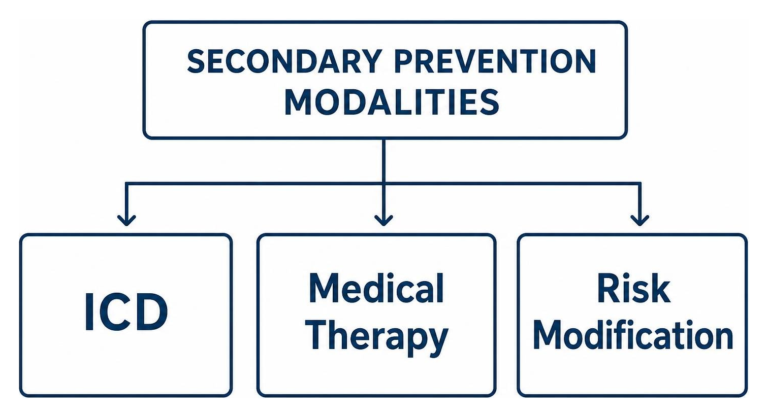
To prevent recurrent sudden cardiac death, patients at elevated risk frequently receive an Implantable Cardioverter-Defibrillator (ICD). Implanted subcutaneously near the collarbone with leads extending into the heart, an ICD continuously monitors the cardiac rhythm 24/7. If it detects life-threatening ventricular fibrillation or tachycardia, it delivers an internal electrical shock to instantly reset the rhythm.
Patients managing cardiovascular conditions can access comprehensive diagnostic heart testing in South Boston, VA through our specialized clinic. If you are seeking specialized long-term cardiac oversight, our team serves as your dedicated South Boston cardiac center.
Frequently Asked Questions about Cardiac Arrest Emergency Treatment
What is the survival rate for out-of-hospital cardiac arrest?
Nationwide, approximately 350,000 Americans experience an out-of-hospital cardiac arrest (OHCA) each year, with an overall survival-to-hospital-discharge rate of roughly 10% (and favorable neurological recovery around 8.2%). In contrast, in-hospital cardiac arrests have a survival rate of approximately 25%. However, when a bystander witnesses the collapse, administers immediate high-quality CPR, and attaches an AED within the first 3 to 5 minutes, survival rates can double or even triple.
Can you harm someone by performing CPR if they are not in arrest?
The risk of causing significant harm by performing chest compressions on someone who is unresponsive is extraordinarily low. While chest compressions can occasionally cause rib fractures or muscular soreness, these injuries heal. Withholding CPR from a person whose heart has stopped is uniformly fatal. If you see someone unresponsive who is not breathing normally, always start CPR.
What should you do if an AED is not immediately available?
If an AED is not nearby, do not delay compressions to search for one. Focus entirely on delivering continuous, high-quality chest compressions at 100 to 120 beats per minute with minimal interruptions. Send another bystander to locate the nearest device while you maintain systemic circulation until EMS personnel arrive with manual defibrillators. If multiple rescuers are present, switch roles every two minutes to prevent rescuer fatigue and maintain compression quality.
Conclusion
When sudden cardiac arrest occurs, the window to act is counted in seconds, not hours. The difference between life and death hinges on ordinary bystanders who recognize the emergency, call 911, push hard and fast on the chest, and deploy an automated external defibrillator before first responders arrive.
Surviving cardiac arrest requires a continuous chain of care—from immediate community CPR and emergency resuscitation protocols to advanced in-hospital interventions, diagnostic testing, and long-term secondary prevention.
At Halifax Heart Center, we are committed to strengthening cardiovascular health and preventing life-threatening cardiac events across South Boston, Halifax County, and Southside Virginia. Whether you are managing an existing cardiovascular condition, seeking preventive screenings, or recovering from a cardiac event, our local cardiology team provides personalized, accessible care. Schedule your consultation for heart disease diagnosis and treatment in South Boston, VA today and take proactive control of your heart health.