
How Cardiology Testing Procedures Find Heart Problems Early
Cardiology testing procedures help doctors identify whether symptoms such as chest pain, unusual fatigue, shortness of breath, or an irregular heartbeat may be caused by a heart problem. Most evaluations begin with low-risk tests, such as blood pressure checks, blood work, an ECG, or an echocardiogram. Depending on your symptoms, health history, and risk factors like high blood pressure, diabetes, smoking, or family history, your doctor may then recommend stress testing, CT or MRI imaging, rhythm monitoring, or an angiogram.
The right test answers a specific question: Is the heart rhythm normal? Are the valves working well? Is blood flow reduced during activity? Is there plaque or a blockage in a coronary artery? Finding the answer early can help guide treatment and lower the risk of serious complications.
At Halifax Heart Center, we provide local, patient-centered care for people throughout South Boston, Halifax County, and Southside Virginia. Our experience with cardiology testing procedures supports accurate diagnosis, early detection, and practical treatment plans tailored to each patient’s needs.
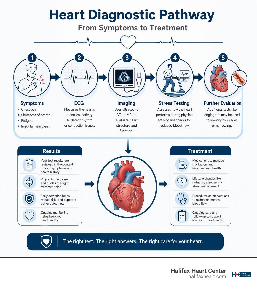
Essential Noninvasive Cardiology Testing Procedures
When evaluating the cardiovascular system, we prioritize noninvasive diagnostics as our primary line of assessment. The heart beats roughly 100,000 times a day—moving blood through thousands of miles of vessels. When something feels off, noninvasive tools give us a look beneath the surface without requiring incisions or hospital stays. These baseline measurements and anatomical evaluations establish what is functioning well and pinpoint any areas requiring closer attention.
Electrical Activity: Electrocardiograms and Ambulatory Monitors
The electrical conduction system of the heart coordinates every contraction. When patients report palpitations, fluttering, dizziness, or unprovoked fatigue, we evaluate this electrical network.
The standard 12-lead electrocardiogram (ECG or EKG) remains one of the most reliable and rapid point-of-care tools in modern medicine. Taking just 5 to 10 minutes, an ECG records electrical impulses through small adhesive electrodes placed on the chest, arms, and legs. It immediately uncovers:
- Baseline rhythm disturbances (such as atrial fibrillation, flutter, or heart blocks)
- Evidence of active or prior myocardial ischemia (reduced blood flow)
- Signs of ventricular hypertrophy (thickening of the heart muscle due to high blood pressure)
However, an in-office ECG is essentially a 10-second snapshot. If an arrhythmia occurs intermittently—such as during sleep, after morning coffee, or during exertional activity—a standard recording may appear completely normal.
To bridge this diagnostic gap, we turn to ambulatory rhythm monitoring. A Holter monitor provides continuous 24- to 48-hour recording of every single heartbeat while you go about your normal daily routine. For symptoms that occur less frequently, such as once a week or once a month, an event recorder or wireless patch monitor can be worn for up to 30 days. These devices are either patient-activated when symptoms occur or automatically triggered by abnormal rhythms. For patients across Halifax County, choosing comprehensive diagnostic heart testing in South Boston, VA ensures these electrical recordings are analyzed quickly by experienced cardiovascular specialists.
Structural Imaging: Echocardiography, Cardiac CT, and Cardiac MRI
While electrical tests show how the heart signals, structural imaging shows how the heart moves, pumps, and holds together.
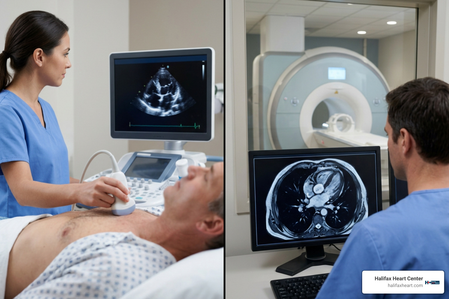
Transthoracic Echocardiogram (TTE): The workhorse of cardiac imaging, the standard echocardiogram uses high-frequency ultrasound waves to create real-time, moving images of the heart’s chambers, muscular walls, and valves. It allows us to calculate the left ventricular ejection fraction (EF)—the percentage of blood pumped out of the main chamber with each beat—and detect valvular regurgitation (leaking) or stenosis (narrowing). For patients requiring high-resolution views of posterior structures, such as when evaluating prosthetic valves or ruling out blood clots in the left atrial appendage, a Transesophageal Echocardiogram (TEE) can be performed under light sedation, placing a specialized ultrasound probe safely down the esophagus.
Coronary Computed Tomography (CT) & Calcium Scoring: Cardiac CT uses specialized X-rays to generate 3D reconstructions of the coronary arteries. A coronary artery calcium (CAC) scan is a non-contrast, 5-to-10-minute scan that measures calcified atherosclerotic plaque in the vessel walls. Expressed as an Agatston score, a result of 0 indicates no identifiable calcified plaque, whereas scores above 100 or 400 indicate moderate to severe atherosclerotic burden, signaling the need for preventive medical therapy. When contrast dye is added, Coronary CT Angiography (CCTA) visualizes the vessel lumen directly, identifying both calcified (hard) and non-calcified (soft) plaques that could obstruct blood flow.
Cardiac Magnetic Resonance Imaging (Cardiac MRI): Cardiac MRI provides clear soft-tissue characterization without utilizing ionizing radiation. Using strong magnetic fields and radio waves, cardiac MRI assesses congenital anomalies, complex cardiomyopathies, pericardial diseases, and viability of the heart muscle. By administering a gadolinium-based contrast agent, we can perform Late Gadolinium Enhancement (LGE) imaging. This technique identifies myocardial scar tissue, fibrosis, and active inflammation (such as myocarditis), clearly distinguishing viable heart muscle from irreversible damage.
Comparing Core Diagnostic Modalities
Selecting the right imaging or electrical test depends on the specific clinical question:
| Diagnostic Modality | Primary Focus | Radiation Exposure | Typical Duration | Best Used For |
|---|---|---|---|---|
| Electrocardiogram (ECG) | Electrical conduction & rhythm | None | 5–10 minutes | Arrhythmia screening, baseline evaluation, acute ischemia detection |
| Transthoracic Echocardiogram | Mechanical function, anatomy & valves | None (Ultrasound) | 30–45 minutes | Heart failure assessment, ejection fraction, valve disorders, pericardial effusion |
| Cardiac Computed Tomography (CT) | Plaque quantification & coronary anatomy | Low to Moderate (Ionizing) | 10–20 minutes | Calcium scoring (CAC), noninvasive coronary stenosis assessment |
| Cardiac Magnetic Resonance (MRI) | Tissue characterization & structural detail | None (Magnetic) | 45–60 minutes | Myocardial viability, scar tissue evaluation, cardiomyopathies, myocarditis |
Evaluating Blood Flow: Stress Tests and Functional Imaging
Coronary artery disease (CAD) often develops quietly. At rest, an artery narrowed by 70% might still deliver enough oxygenated blood to prevent symptoms. But when physical exertion increases oxygen demand, that narrowed vessel fails to keep pace, triggering myocardial ischemia (oxygen deprivation to the muscle). Cardiac stress testing unmasks these exertional limitations.
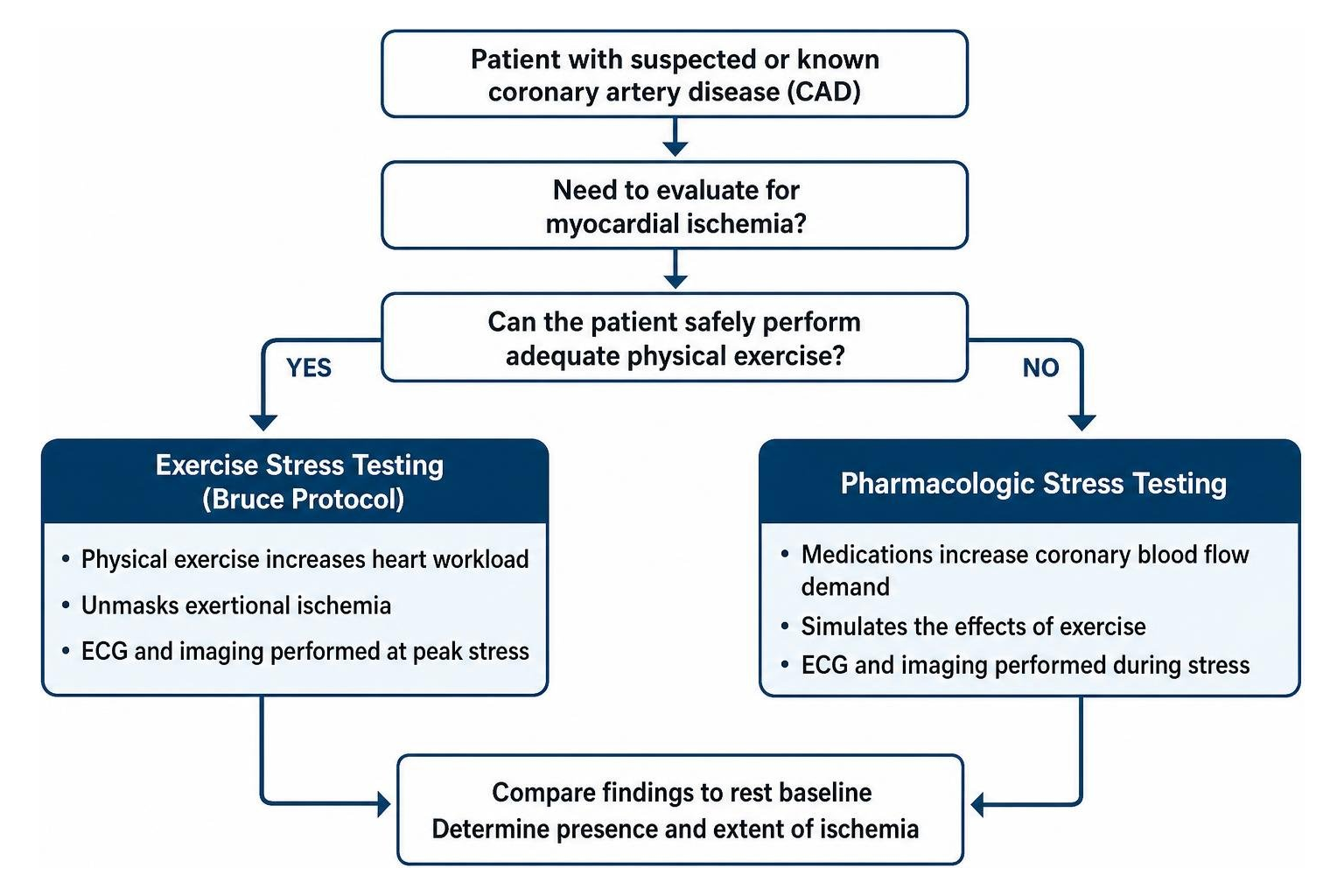
Exercise vs Pharmacologic Stress Protocols
Stress testing can be performed using physical exercise or targeted medications to increase the heart’s workload in a safe, controlled setting.
- Exercise Stress Testing (The Bruce Protocol): When a patient is physically capable of walking, exercise testing on a treadmill is the preferred modality because it reproduces real-world physiological demand. Under the standard Bruce protocol, the speed and elevation of the treadmill increase every 3 minutes. Our clinical team continuously monitors your 12-lead ECG, blood pressure, heart rate, and physical symptoms. The primary objective is to reach your target heart rate—typically 85% of your age-predicted maximum (calculated as 220 minus your age)—while monitoring for exercise capacity, rhythm changes, or ST-segment depressions that signal ischemia.
- Pharmacologic Stress Protocols:
For patients with severe arthritis, peripheral vascular disease, mobility limitations, or chronic obstructive lung conditions that prevent sustained treadmill exercise, medications simulate the effects of exercise:
- Vasodilators (Adenosine, Dipyridamole, Regadenoson): These agents selectively dilate normal coronary arteries. Diseased vessels with fixed atherosclerotic narrowings cannot dilate to the same degree, creating a relative blood flow deficit that imaging detects.
- Inotropic Agents (Dobutamine): For patients who cannot exercise and have contraindications to vasodilators (such as severe reactive airway disease or asthma), dobutamine is infused incrementally to increase heart rate and myocardial contractility, mimicking physical exercise.
Our clinical staff reviews all relative and absolute contraindications prior to testing. Stress tests are avoided in situations involving acute coronary syndromes, severe symptomatic aortic stenosis, uncontrolled arrhythmias, acute pulmonary embolism, or decompensated heart failure, adhering closely to recognized cardiac stress testing protocols and safety standards.
Advanced Stress Cardiology Testing Procedures
A standard treadmill stress test evaluating ECG changes alone provides valuable baseline information, yielding a diagnostic sensitivity for CAD of 58% to 66% and a specificity of 61% to 62%. However, pairing physical or pharmacologic stress with advanced imaging raises diagnostic accuracy.
Stress Echocardiography: Ultrasound images of the heart are captured at rest and immediately following peak exercise (or during dobutamine infusion). If a coronary artery has a flow-limiting blockage, the segment of heart muscle supplied by that vessel will fail to contract normally under stress, developing a transient wall motion abnormality. Stress echocardiography raises diagnostic sensitivity to 81%–85% and specificity to 82%–85%.
Radionuclide Myocardial Perfusion Imaging (SPECT and PET): Nuclear stress testing uses small, safe amounts of radioactive tracers (such as Technetium-99m sestamibi or Rubidium-82) injected intravenously (Gopal et al., 2025). Single-Photon Emission Computed Tomography (SPECT) and Positron Emission Tomography (PET) cameras record tracer uptake within the heart muscle at rest and during peak stress.
- Reversible Ischemia: A region showing normal tracer uptake at rest but reduced uptake during stress indicates an area of muscle that is viable but suffering from reduced blood flow due to a narrowed artery.
- Fixed Defect: Reduced tracer uptake during both rest and stress indicates prior infarction and permanent scar tissue.
Radionuclide myocardial perfusion imaging provides a diagnostic sensitivity of 82% to 92% and specificity of 70% to 81%, making nuclear medicine stress testing methods a reliable tool for risk stratification.
Stress Cardiac MRI: Stress perfusion MRI combines pharmacologic stress with real-time gadolinium imaging, yielding a sensitivity of 83% to 87% and a specificity of 83% to 93% without any exposure to ionizing radiation.
Invasive Diagnostics: Cardiac Catheterization and Angiography
When noninvasive testing indicates significant ischemia, or when a patient presents with unstable symptoms, invasive diagnostics provide definitive anatomical visualization and hemodynamic measurement.
Coronary Angiography as the Gold Standard
Coronary angiography (performed during cardiac catheterization) remains the clinical gold standard for identifying the presence, location, and severity of coronary artery disease.
During the procedure:
- A small sheath is introduced into an artery—most commonly the radial artery in the wrist, or alternatively the femoral artery in the groin. Radial access significantly reduces access-site bleeding complications, lowers hematoma risk, and allows patients to sit up and walk shortly after the procedure.
- A long, thin, flexible catheter is guided under X-ray imaging (fluoroscopy) to the origins of the coronary arteries.
- A small volume of radiopaque contrast dye is injected directly into the coronary vessels while high-resolution, moving X-ray images are recorded.
Angiography reveals the exact percentage of coronary artery stenosis (narrowing). If a severe, flow-limiting blockage is identified, interventional cardiologists can often transition immediately from diagnosis to treatment, performing balloon angioplasty and deploying a drug-eluting stent to restore coronary blood flow. Learning more about cardiac catheterization procedures and risks helps patients understand how diagnostic insight and therapeutic relief can occur during the same clinical visit. For those navigating complex heart concerns, timely heart disease diagnosis and treatment in South Boston, VA provides access to these advanced diagnostic and interventional pathways close to home.
Specialized Invasive Cardiology Testing Procedures
Modern catheterization labs go beyond standard visual imaging. Visual assessments alone can sometimes struggle to determine whether an intermediate narrowing (such as a 40% to 60% blockage) is truly restricting blood flow to the heart muscle. Advanced physiology and intravascular imaging tools resolve these uncertainties:
- Fractional Flow Reserve (FFR) & Non-Hyperemic Pressure Ratios (iFR/NHPR): A specialized, pressure-sensing guidewire is advanced past a coronary lesion. By comparing blood pressure distal to the blockage against the pressure in the aorta, we calculate a precise ratio. An FFR value of ≤ 0.80 or an NHPR value of ≤ 0.89 confirms that the lesion is hemodynamically significant and would benefit from stenting, whereas higher values indicate the blockage can be safely managed with medication.
- Intravascular Ultrasound (IVUS) & Optical Coherence Tomography (OCT): These miniaturized imaging probes are threaded directly inside the coronary artery. IVUS uses sound waves and OCT uses light waves to generate cross-sectional images from inside the vessel. They reveal the true thickness of the plaque, identify vulnerable lipid pools or calcifications, and confirm that an expanded stent is well-apposed against the vessel wall.
- Hemodynamic Monitoring & Endomyocardial Biopsy: During right heart catheterization, pressures within the right atrium, right ventricle, pulmonary artery, and pulmonary capillary wedge position are measured to diagnose pulmonary hypertension and assess complex heart failure. When rare conditions like amyloidosis, sarcoidosis, or acute myocarditis are suspected, small tissue samples can be safely collected via endomyocardial biopsy. These advanced cardiac catheterization techniques provide comprehensive diagnostic clarity when noninvasive findings remain inconclusive.
Blood Biomarkers and Clinical Decision-Making
High-tech imaging provides structural and functional insight, but laboratory blood tests provide crucial molecular and biochemical context. Blood biomarkers uncover active myocardial injury, underlying metabolic risk, and systemic vascular inflammation.
Key Laboratory Markers: Lipid Panels, Troponin, and CRP
- High-Sensitivity Cardiac Troponin (hs-cTnI / hs-cTnT): Troponins are regulatory proteins found exclusively within cardiac muscle cells. When heart muscle cells experience acute injury or oxygen starvation, troponin leaks into the bloodstream. High-sensitivity troponin assays can detect even microscopic levels of myocardial necrosis within hours, serving as the essential laboratory test for ruling in or ruling out an acute myocardial infarction (heart attack).
- Lipid Profile & Apolipoproteins: A standard lipid panel measures total cholesterol, triglycerides, High-Density Lipoprotein (HDL), and Low-Density Lipoprotein (LDL). Elevated LDL cholesterol directly contributes to the development and progression of arterial plaque. Advanced lipid panels may also evaluate Apolipoprotein B (ApoB) and Lipoprotein(a), which provide a more detailed assessment of inherited cardiovascular risk.
- High-Sensitivity C-Reactive Protein (hs-CRP): Atherosclerosis is an active inflammatory process. Elevated hs-CRP levels reflect systemic vascular inflammation, helping stratify cardiovascular risk in patients who have borderline cholesterol levels.
- B-Type Natriuretic Peptide (BNP & NT-proBNP): When the heart chambers experience increased stretch, pressure, or volume overload, the heart muscle releases BNP. Measuring BNP levels is invaluable for diagnosing and monitoring heart failure, as well as distinguishing cardiac causes of shortness of breath from pulmonary conditions like asthma or COPD.
How Cardiologists Select the Right Diagnostic Pathway
Cardiologists do not order tests at random. We use an evidence-based approach guided by your individual pre-test probability—the likelihood that your symptoms are caused by coronary artery disease based on your age, sex, symptom description (typical angina vs. atypical chest discomfort), and baseline risk factors.
Reviewing our complete guide to cardiovascular testing and consults illustrates how clinical guidelines help us choose the most direct, comfortable, and accurate diagnostic pathway for each patient.
Post-Test Care: Treatment Plans and Lifestyle Adjustments
A cardiac test result is never an endpoint—it is the roadmap that guides your care. Once diagnostic procedures are complete, our clinical team works alongside you to interpret the results and build a personalized treatment strategy.
If testing reveals normal findings, care shifts toward primary prevention, maintaining healthy lifestyle habits, and scheduling routine checkups. If tests reveal evidence of cardiovascular disease, our management strategies include:
- Evidence-Based Medical Therapy: Starting guideline-directed medications such as statins (to lower LDL and stabilize arterial plaque), antiplatelet therapy (like aspirin), beta-blockers (to reduce heart rate and blood pressure), or ACE inhibitors/ARBs/ARNIs (to protect heart muscle and kidney function).
- Interventional Revascularization: When severe blockages are identified, procedures such as percutaneous coronary intervention (PCI) with stent placement or a referral for coronary artery bypass graft (CABG) surgery may be recommended to restore normal blood flow.
- Structured Cardiac Rehabilitation: A medically supervised exercise and educational program designed to strengthen cardiovascular endurance, improve functional capacity, and support long-term recovery following a cardiac event or stent placement.
- Heart-Healthy Lifestyle Adjustments: Sustainable daily changes remain the foundation of cardiovascular health. This includes adopting a Mediterranean-style or low-sodium dietary pattern, engaging in 150 minutes of moderate-intensity aerobic exercise each week, achieving restorative sleep, managing daily stress, and stopping all tobacco and nicotine use.
Through our comprehensive heart services, we provide continuous support, ongoing monitoring, and timely treatment adjustments to help you maintain optimal cardiovascular wellness.
Frequently Asked Questions About Cardiac Diagnostics
How do doctors decide which heart test is right for me?
We evaluate your specific symptoms (such as whether chest pressure occurs with exertion or at rest), your personal medical history, your physical exam findings, and your calculated cardiovascular risk score. We typically start with noninvasive, low-risk tests—such as an ECG, standard blood panels, or an echocardiogram—before advancing to stress imaging, CT angiography, or invasive cardiac catheterization.
What preparation is required before cardiac CT or MRI imaging?
For a cardiac CT or calcium score, you may be asked to fast for 3 to 4 hours prior to the scan and avoid caffeine, nicotine, and energy drinks for 12 to 24 hours, as elevated resting heart rates can blur CT images. If contrast dye is planned, we may run a quick blood test to verify normal kidney function. For a cardiac MRI, you must inform our team of any implanted medical devices, metal fragments, or claustrophobia so we can make appropriate safety adjustments.
What are the primary risks associated with cardiac stress testing?
Cardiac stress testing is performed under continuous clinical supervision. Serious complications, such as a heart attack or severe arrhythmia, are rare, occurring in approximately 0.06% of patients tested. If pharmacologic agents like dipyridamole or regadenoson are used, temporary side effects such as flushing, headache, shortness of breath, or mild chest discomfort can occur in 10% to 20% of patients, but these symptoms typically resolve within minutes or can be quickly reversed with medications like aminophylline.
Conclusion
Cardiovascular disease remains the leading health challenge across the nation, but modern cardiology testing procedures provide the clarity needed to detect, manage, and prevent serious complications before they disrupt your life. From a 10-minute ECG and detailed ultrasound imaging to advanced nuclear stress testing and high-precision coronary angiography, today’s diagnostic tools take the guesswork out of heart health.
If you are experiencing unexplained chest discomfort, shortness of breath, palpitations, or simply want to understand your personal cardiovascular risk, do not wait for symptoms to worsen. Contact our dedicated team at Halifax Heart Center to schedule a personalized cardiac consultation at our South Boston clinic today.
Scientific References
Shwetha Gopal, Thomas F. Heston, Christie Murphy. “Nuclear Medicine Stress Test.” StatPearls Publishing, 2025. NBK557682.